ABSTRACT
In our previous study, artificial intelligence technique has been applied for analysis of FT-IR spectra of human blood plasma samples to classify carcinoma and healthy
people. Various statistical methods, such as “Linear SVM” (support vector machine), “PLS-DA” (partially square discriminant analysis) and “Random Forest” were used for
classification to distinguish a group of patients and healthy people. Prediction accuracies were 90% and 80%, respectively. In this study, the increase molecular features
that differentiate healthy and cancer patients, FT-IR spectra of both plasma and its lipid fractions were analyzed. Numerous spectral peaks of lipid fraction are not seen
in blood plasma FT-IR spectra. Functional groups identified from FT-IR spectra of the lipid fraction of blood plasma make valuable impact for accurate classification. In
the study, blood plasma samples were collected from 50 patients and 49 healthy individuals. Then, lipids were extracted for each plasma sample. Coupled analysis was
performed for FT-IR spectra of the plasma and its corresponding lipid fractions. For both groups the greatest differences in plasma and lipid fractions FT-IR spectra were
observed around 3280 sm-1, 3060 sm-1, 2960 sm-1, 2930 sm-1, 1245 sm-1 and 3016 sm-1, 2920 sm-1,
1650 sm-1, 1214 sm-1, respectively. The data remaining outside the standard deviations in the infrared spectra of plasma and lipid samples allow it to be
used as a preliminary diagnostic marker in determining the disease. The work in progress to apply artificial intelligence technique for detailed classification.
Keywords: lung adenocarcinoma, blood plasma, blood plasma lipids, FCHIG spectroscopy.
PACS: 87.64.Ve
DOI:-
Received: 09.12.2022
AUTHORS & AFFILIATIONS
1. Institute of Biophysics, Ministry of Science and Education Republic of Azerbaijan 117 Z. Khalilov, Baku, AZ 1141
2. National Oncology Center of the Ministry of Health of the Republic of Azerbaijan, 79B H. Zardabi Street
E-mail: oktaygasimov@gmail.com
Graphics and Images
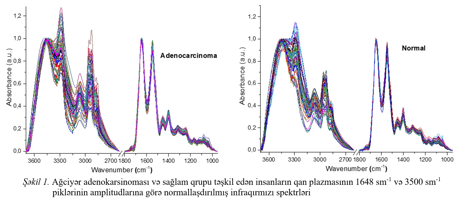
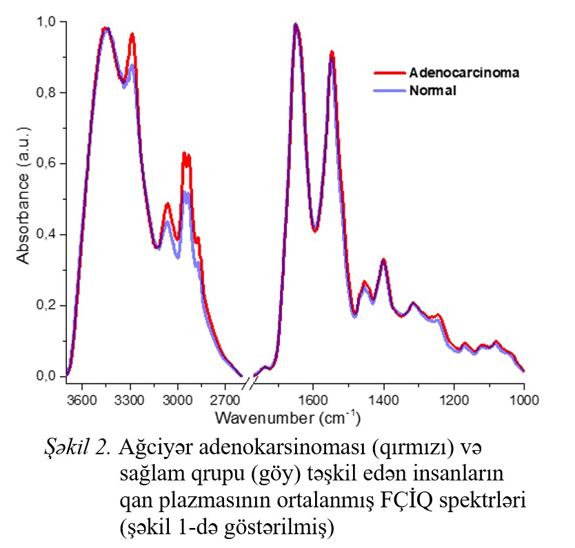
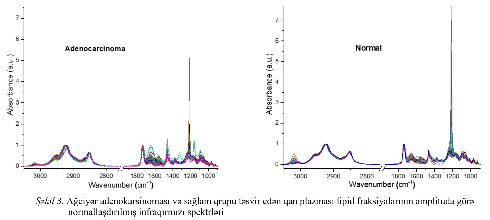
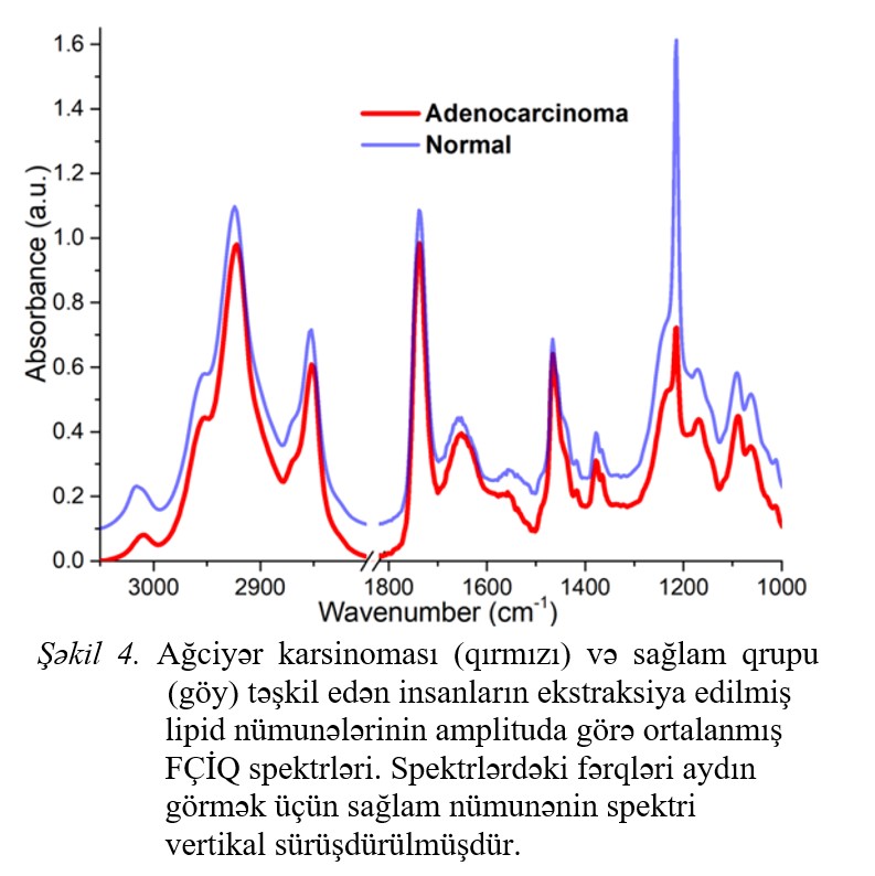
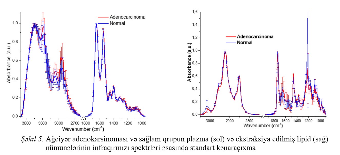
Fig.1 Fig.2 Fig.3 Fig.4 Fig.5
|
REFERENCIES
[1] J.Ferlay, C.Partensky, F.Bray. More deaths from pancreatic cancer than breast cancer in the EU by 2017. Acta Oncologica, 2016, vol.55 Issue 9-10, pp.1157-1160.
[2] J. Ferlay, Isabelle Soerjomataram. Cancer incidence and mortality worldwide: sources, methods and major patterns in Globocan 2012, 2015, vol.136, Issue 5, pp.359-386.
[3] Rebecca L. Siegel, Stacey A.Fedawa. Colorectal cancer incidence patterns in the United States 1974-2013, 2017, vol. 109, Issue 8, pp.1-6.
[4] O.K.Gasymov, A.H.Aydemirova, L.A.Melikova, J.A.Aliyev.App.Comp.Math.,vol.20.№ 2.pp.277-289.
[5] M.J. Thun et al Journal of the National Cancer Institute, vol.98, № 19, 2006, pp.691-699.
[6] Basil Rigas., Patrick T.T.Wong. Human colon adenocarcinoma cell line display infrared spectroscopic features of malignant colon tissues, Cancer Research, 1992, vol.52, pp.84-88.
[7] H.P.Wang, H-C Wang, Y-J Huang. Microscopic FTIR studies of lung cancer cells in pleural fluid. The science of the total environment., 1997, vol.204. pp. 283-287.
[8] A.H. Aydəmirova. 2022, AJP FİZİKA, vol. XXVIII, Issue 2, pp.12-16.
[9] Kan-Zhi Liu, Kam Sze Tsang, Chi Kong Li. Infrared spectroscopic identification of betta-thalassemia., Clinical chemistry, 2003, Vol.49 (7), pp.1125-1132.
[10] Anila Kumari, Shalmali Bhattacharya. Application of FTIR spectroscopy as a tool for early cancer detection. American Journal of biomedical sciences. 2018, ISSN (1937-9080), pp. 139-48.
[11] Daping Sheng, Yican Wu, Xin Wang. Comparison of serum from gastric cancer patients and from healthy persons using FTIR spectroscopy. Spectrochimica Acta-Part A, 2013(116), pp.365-369.
[12] Janina Kneipp, Michael Beekes., Molecular changes of preclinical scrapie can be detected by infrared spectroscopy. The Journal of Neuroscience, 2002., vol 22(8), pp. 2989-2997.
[13] Zanyar M., Shazza R.&Dr.Intesham ur Rehman. Fouries Transform Infrared (FTIR) spectroscopy of biological tissues. Applied Spectroscopy Reviews, vol.13, Issue 2, February 2008, pp.134-179.
[14] Abasalt Hosseinzadeh Colagar et al. Fourier transform infrared miscrospectroscopy as a diagnostic tool for distinguishing between normal and malignant gastric tissue. J.Biosci, 2011, vol.36, Issue 4, pp.669-677.
[15] Evgeny Bogomolny, Shmuel Argov. Monitoring of viral cancer progression using FTIR microscopy. A comparative study of intact cells and tissues; Bopchimica et Biophysica Acta. 2008, vol.1780, Issue 9, pp.1038-1046.
[16] Lukas K.Tamm, Suren A.Tatulian. Infrared spectroscopy of proteins and peptides in lipid bilayers, QRB, 1997, vol.30, Issue 4, pp.365-429. John H.Crowe, Folkert A.Hoekstra. Lipid phase transitions measured in intact cells with Fourier transform infrared spectroscopy. Crybiology, 1989, vol.26, Issue 1, pp.76-84.
[17] E.G. Bligh, W.J.Dyer. Canadian Journal of Biochemistry and Physiology, vol.37, Issue 8; pp.911-917.
[18] Jiansheng Wang, Jia Zhang et al. Evaluation of gallbladder lipid level during carcinogenetics by an infrared spectroscopic method. Original article. 2010, vol.55, pp.2670-2675.
[19] Sumanta Kar, Dinesh R.Katti et al. Spectrochimica Acta Part A, Molecular and biomolecular spectroscopy, 2019; vol.208, pp.85-96.
[20] Michael Fung Kee Fung, Mary K.Senterman et al. Biospectroscopy, 1996, vol.2, Issue 3, pp.155-165.
[21] P.Venkatachalam, L.Lakshmana Rao et al. Dignosis of breast cancer based on FT-IR spectroscopy; AIP Conference Proceedings; 2008, vol.1075, Issue 144, pp.144-148.
[22] V.Crupi, D.De Domenico et al. Journal of Molecular Structure, 2001, vol.563-564, pp.115-118.
[23] S.B. Akkas, M. Severcan et al. Effects of lipoic acid supplementation on rat brain tissue, An FT-IR spectroscopic and neural network study, 2007, vol.105, Issue 3, pp.1281-1288.
[24] Isabell Dreissig, Susane Machill et al. Quantification of brain lipids by FT-IR spectroscopy and partial least squares regression, Spectrochimica Acta Part A. Molecular and Biomolecular Spectroscopy, 2009, vol.71, Issue 5, pp. 2069-2075.
[25] Maria Paraskevaidi, Camilo L.M. Morais et al. Detecting endometrial cancer by blood spectroscopy a diagnostic cross-sectional study; Cancers, 2020, vol.12, Issue 5, pp1-17.
|
